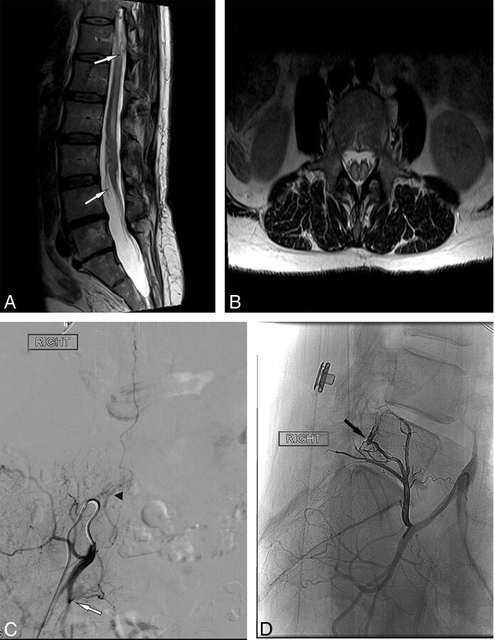
Fig 3.
A, Sagittal T2-weighted MR imaging demonstrates tethering of the spinal cord with the tip of the conus at the L3–4 interspace (white arrow) before surgical release. There is high T2 signal of the cord and a faintly visualized flow void (upper white arrow). B, Axial T2-weighted MR imaging shows partial diastematomyelia in the lumbar spine without evidence of a fibrous band or boney spur/bar. High cord signal is present in both hemicords. C, Right internal iliac artery spinal angiogram demonstrates an epidural fistula arising from the right lateral sacral artery (white arrow) with an intradural draining vein (arrowhead). D, Post-Onyx embolization angiogram demonstrates an Onyx cast in the ventral epidural space of L5 (black arrow).

