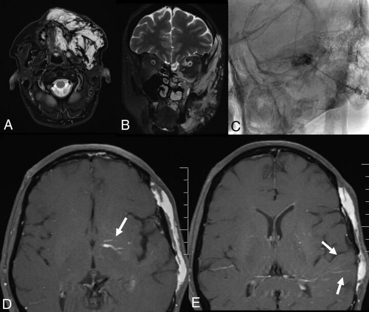
Fig 1.
A 49-year-old woman with VMs and DVAs. VMs are in the left temporal region, orbit, zygomaticofacial region, and mandible (A and B). The patient underwent bleomycin sclerotherapy for treatment of the VMs with good results (C). She also had an extensive DVA of the left temporal lobe, basal ganglia, and left cerebellar hemisphere (D and E). Findings would be consistent with CVMS 1–3.

