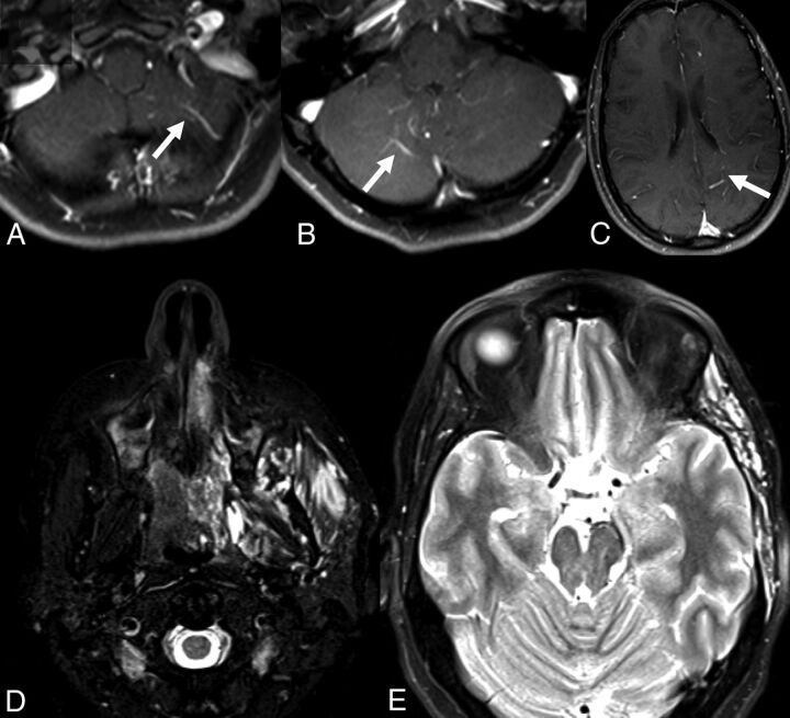
Fig 2.
A 30-year-old man with facial VMs and left supratentorial DVAs. A–C, Postcontrast T1-weighted MRIs show DVAs in the bilateral cerebellar hemispheres, left parietal lobe, and left temporal lobe. D and E, T2-weighted MR imaging shows extensive VMs of the left zygomaticotemporal region and masticator spaces.

