Fig 3.
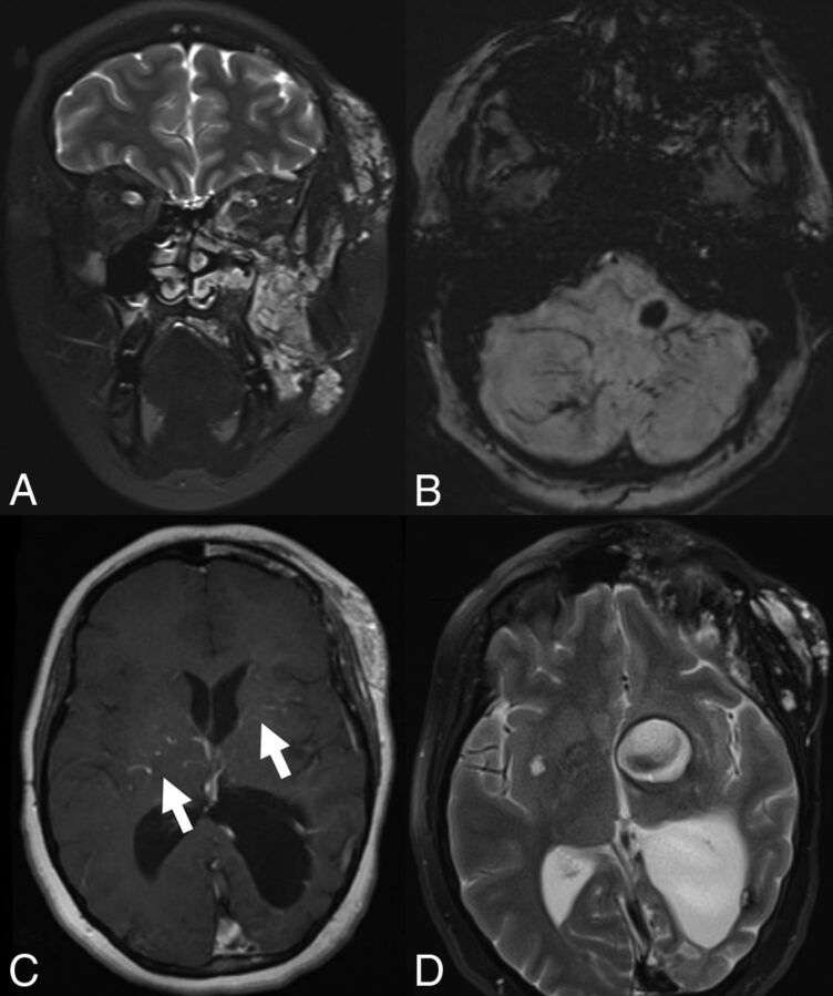
A 22-year-old woman with a left facial DVA, bilateral VMs, and a de novo cavernoma. A, Coronal T2-weighted MR imaging demonstrates a vascular malformation involving the soft tissues overlying the left zygomatico-orbital region with extension to the left maxillary region. B, Axial SWI MR imaging of the brain demonstrates extensive DVAs of the bilateral cerebellar hemispheres and a cavernoma of the left medulla. C, Postcontrast MR imaging demonstrates venous radicles of 2 DVAs involving the bilateral basal ganglia. D, T2-weighted MR imaging 2 years later demonstrates a large cavernoma that developed in one of the venous radicles of the left basal ganglia DVA.

