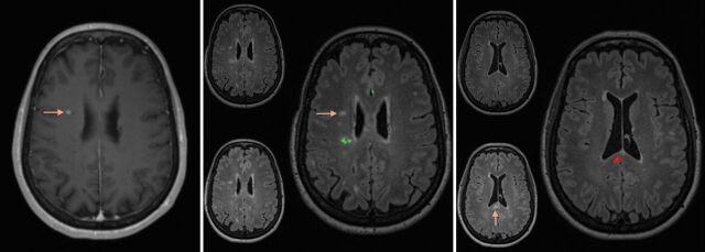
Fig 2.
Left: Contrast-enhanced T1 MR imaging shows enhancement of a pre-existing lesion in the right centrum semiovale (arrow). Middle: The composite image from the coregistering software shows no growth of this corresponding lesion on FLAIR between the prior and current study (arrow). Additionally, 1 lesion had slightly regressed in the right posterior corona radiata (green on the composite image). Right: The composite image using the coregistering software at a more inferior level shows evidence of a new lesion in the splenium of the corpus callosum (arrow and red on composite image on the right).

