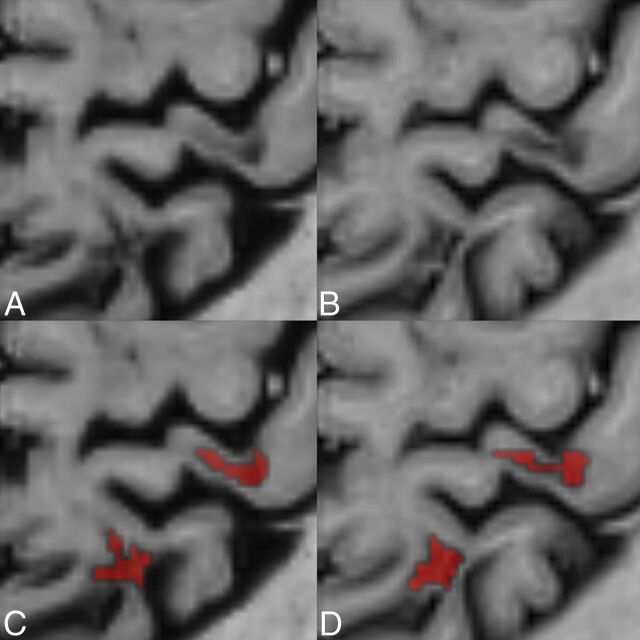
Fig 2.
Comparison between conventional and synthetic phase-sensitive inversion recovery. A comparison between conventional (B and D) and synthetic phase-sensitive inversion recovery (A and C) illustrates 2 leukocortical MS lesions in a 40-year-old female patient with MS. Lower row illustrates the manual segmentation of the lesions by a neuroradiologist.

