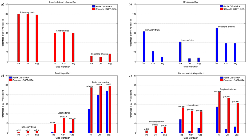
Figure 2: Evaluating the specific and general image artifacts in healthy volunteers (HV).
The percentage of occurrence of each imaging artifact is demonstrated as a function of slice orientation in three different pulmonary regions. The diagram is based on the results of the two readers together. The 60 datasets comprised 30 independently evaluated datasets of each radiologist.

