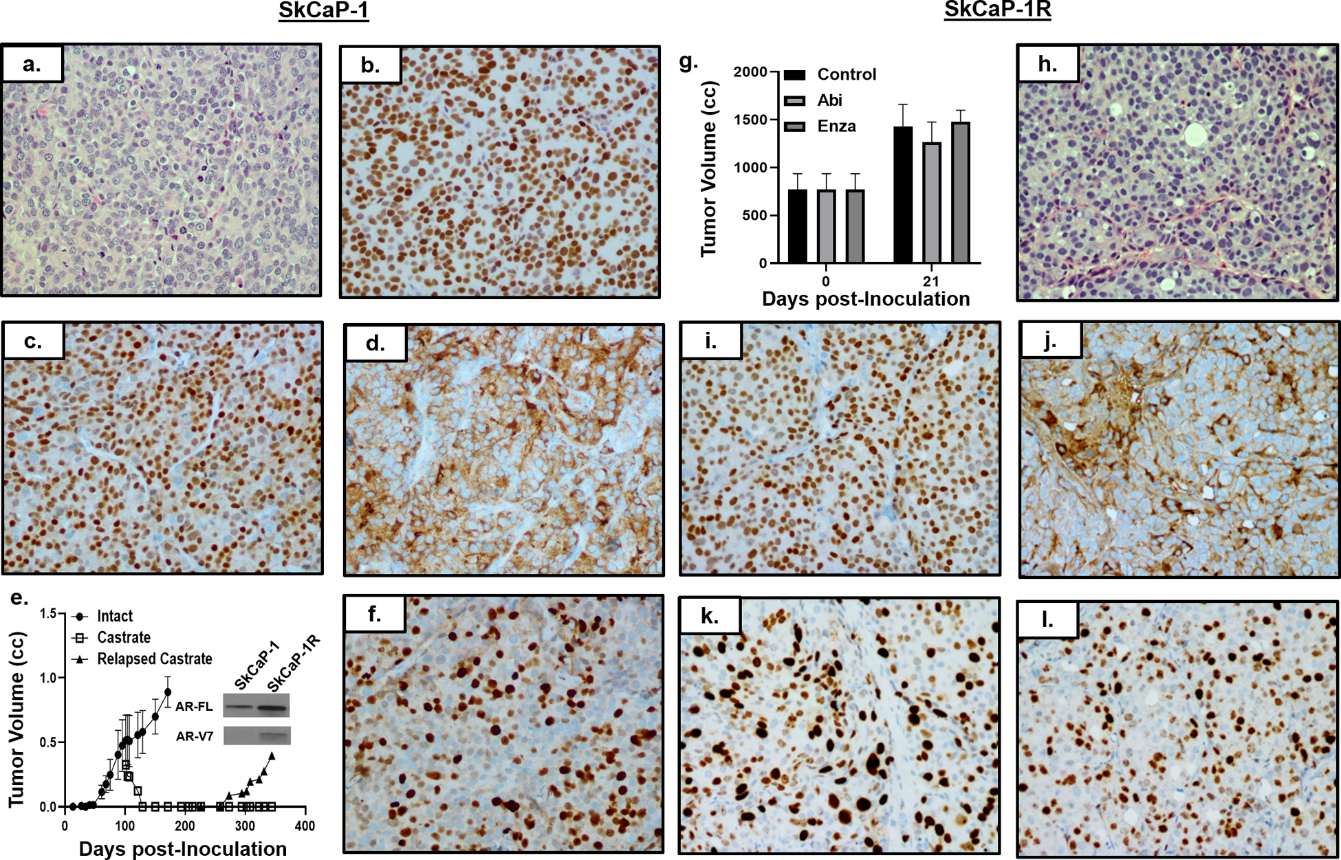
Figure 4: Characterization of SkCaP-1 and SkCaP-1R.

a) H & E histology (200x) of SkCaP-1. IHC (200x) of SKCaP-1 for b) AR, c) Nkx3.1, and d) PSMA. e) Growth rate of SkCaP-1 in intact (i.e. ADT-equivalent) mice with subsequent regression and relapse in castrate (i.e. ARSi-equivalent) male NSG mice (n = 5 each). AR-FL and AR-V7 immunoblots of SkCaP-1 vs. SkCaP-1R (inset). f) IHC (200x) of SkCaP-1 for Ki67. g) Abi and Enza resistance of SkCaP-1R in vivo (n = 3 each). h) H & E histology (200x) of SkCaP-1R. IHC (200x) of i) AR, j) PSA, k) c-Myc, and i) Ki67 in SkCaP-1R PDX.
