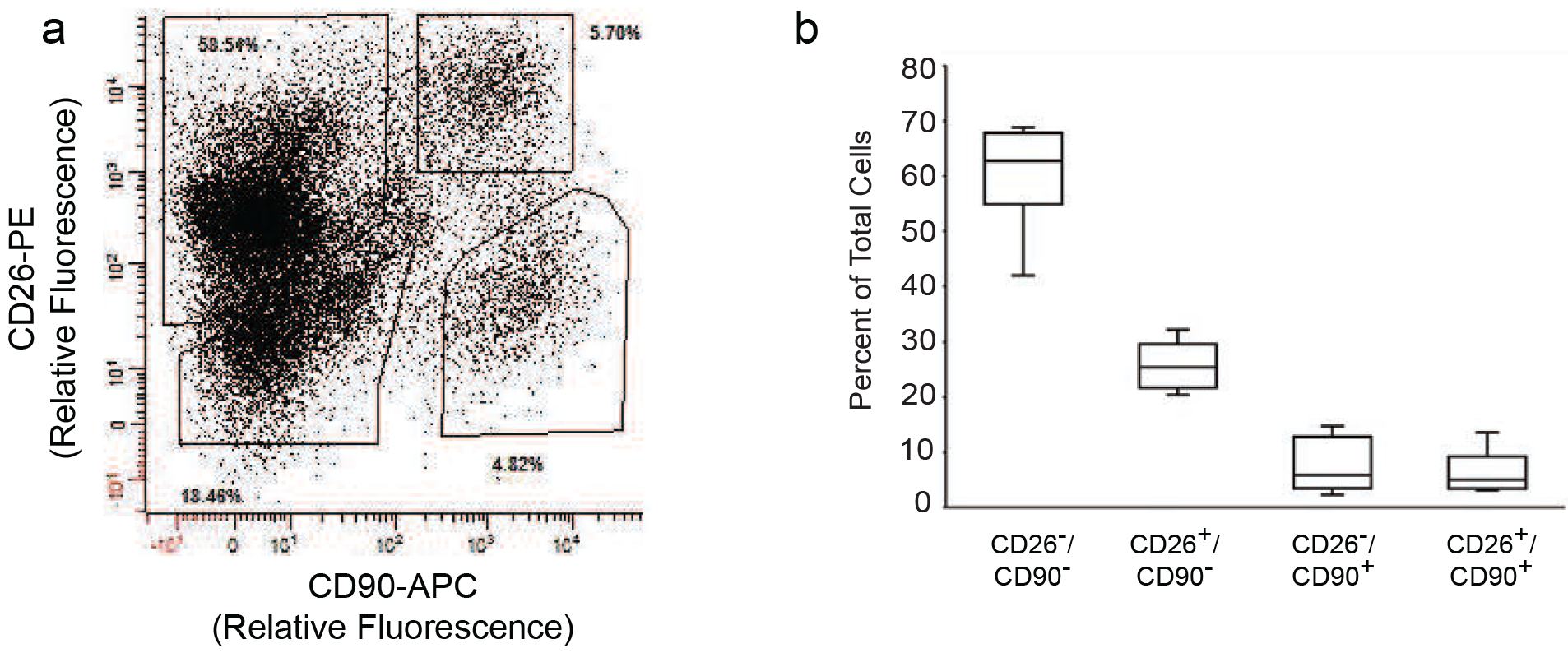
Figure 1. Identification of CD26+ cell populations in human dermis.

Dermal rich skin cell suspensions were prepared by collagenase digestion of adult human buttocks skin samples. Cells were labeled with fluorophore-conjugated antibodies to CD26, CD90, CD34 and CD45, followed by four-color flow cytometry analysis. A) Representative flow cytometry analysis showing distribution of cell populations based on expression of CD26 and CD90. The solid polygons depict gates that were used for sorting. Percentages of total cells within each gate are indicated. B) Quantitation of CD26 and CD90 cell populations in adult human dermis. Results are means+SEM, n=5 subjects.
