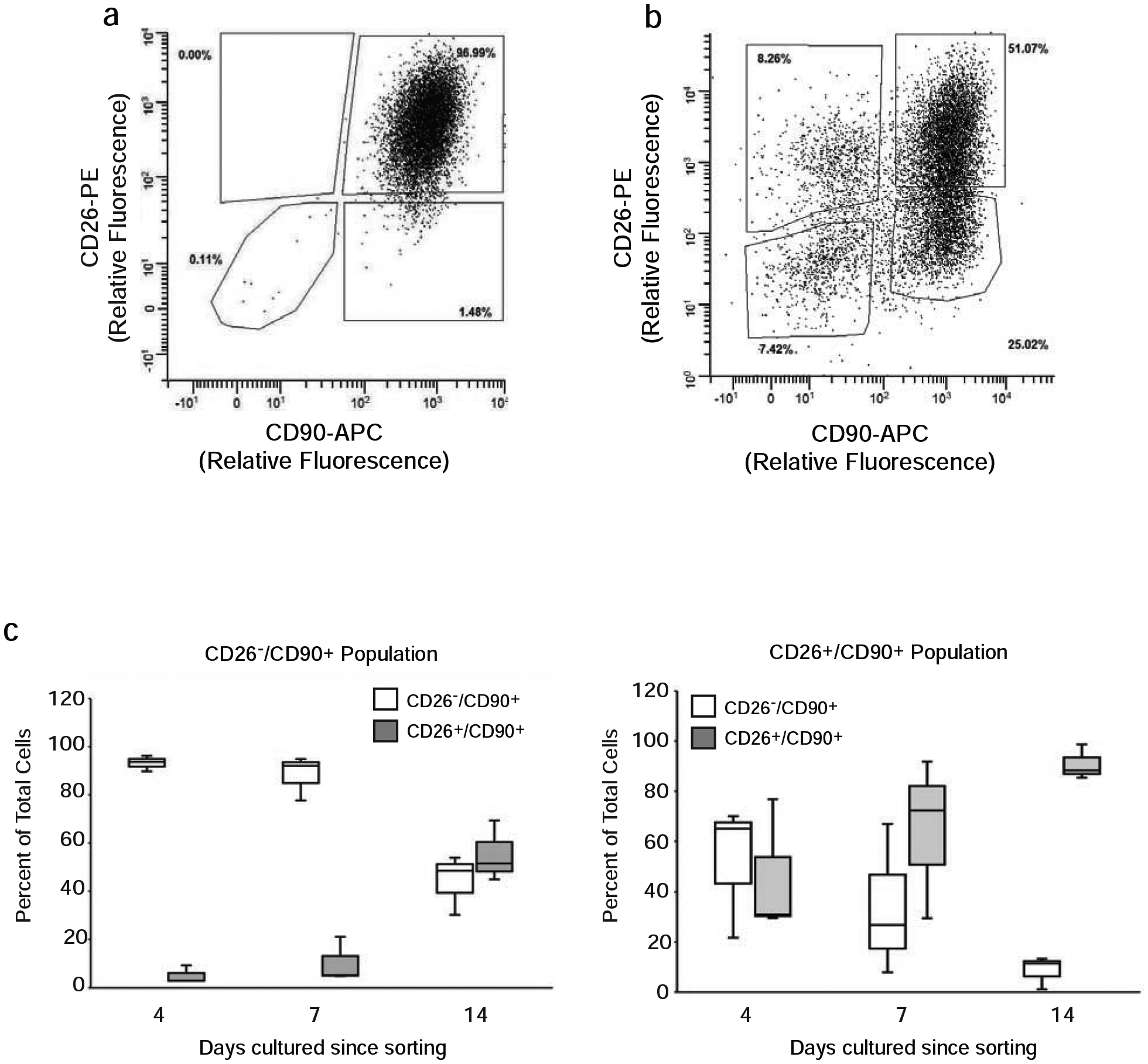
Figure 5. Primary dermal fibroblasts express CD26 in culture.

A) Dermal cells from buttocks skin were grown in culture through three passages and then analyzed by flow cytometry as described in the Figure 1 legend. Results are representative of five subjects. B) Freshly isolated dermal cells from buttocks skin were placed in culture for three days, and adherent cells were analyzed and sorted by FACS, as described in Figure 2 legend. Results are representative of five subjects. C) Following FACS sorting as in (B), 105 CD26− and CD26+ fibroblasts (CD90+) were separately cultured. Cultures were analyzed for CD26 and CD90, at the indicated times in culture by flow cytometry. Graphs are the average of five different subjects.
