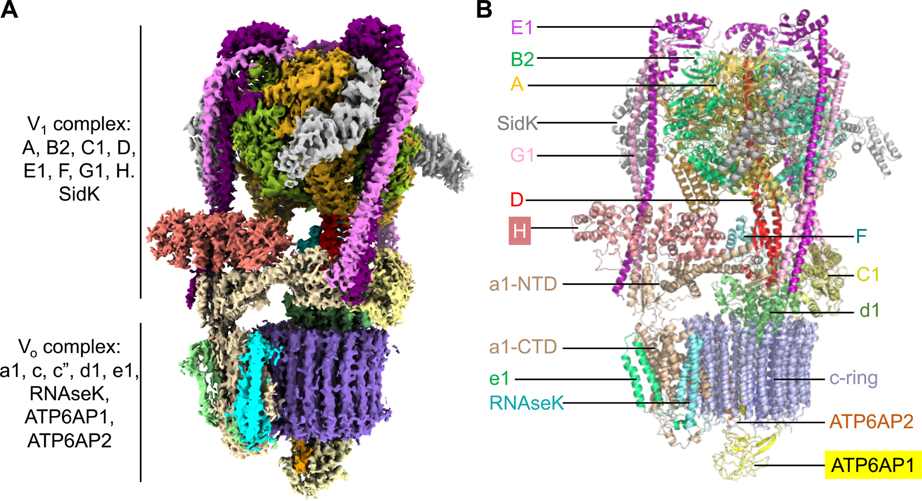
Figure 1. Cryo-EM Structure of Human V-ATPase.

(A) Cryo-EM density of human V-ATPase (state 1) with subunits color coded.
(B) Ribbon diagram of human V-ATPase structure (state 1) with subunits color coded and labeled. Subunits H and ATP6AP1 which are absent or not fully traced in the rat V-ATPase structure (15) are highlighted.
See also Table S1 and Figures S1 and S2.
