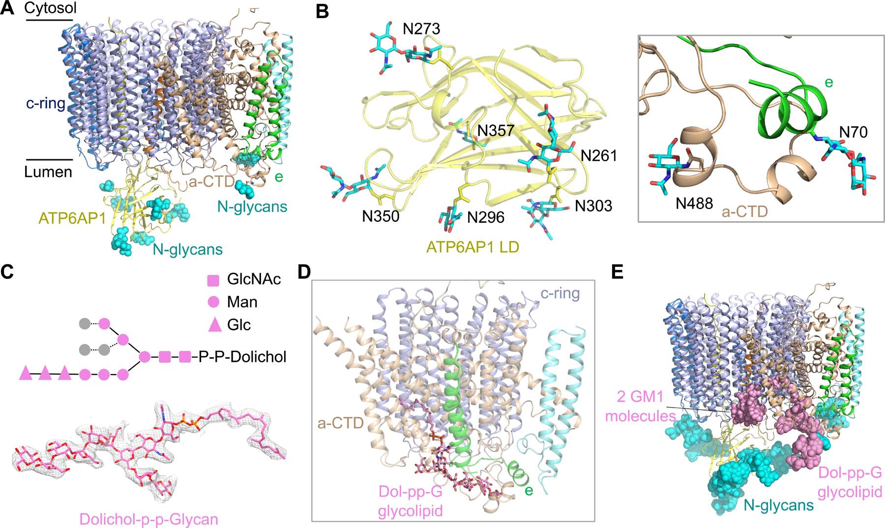
Figure 5. N-linked Glycosylation and Glycolipids of the Vo Complex.

(A) N-linked glycans (cyan spheres) at the luminal side of the Vo complex from subunits ATP6AP1, a-CTD and e.
(B) Detailed structures of N-linked glycan units on subunits ATP6AP1, a-CTD and e.
(C) Diagram of the chemical structure, the model (in stick) and the cryo-EM density (2.0 σ) of the glycolipid dolichol-P-P-glycan (Dol-pp-G). Two copies of N-acetylglucosamine (GlcNAc) (square), six mannose (circle), and three glucose (triangle) units were resolved in the cryo-EM density map.
(D) Dol-pp-G bound to subunits c, a-CTD and e.
(E) The Vo complex shown with observed glycans at the luminal side. For each N-linked glycosylation site, a total of nine sugar units were modeled based on the complex-type N-glycan structure. GM1: monosialoganglioside.
See also Figure S5.
