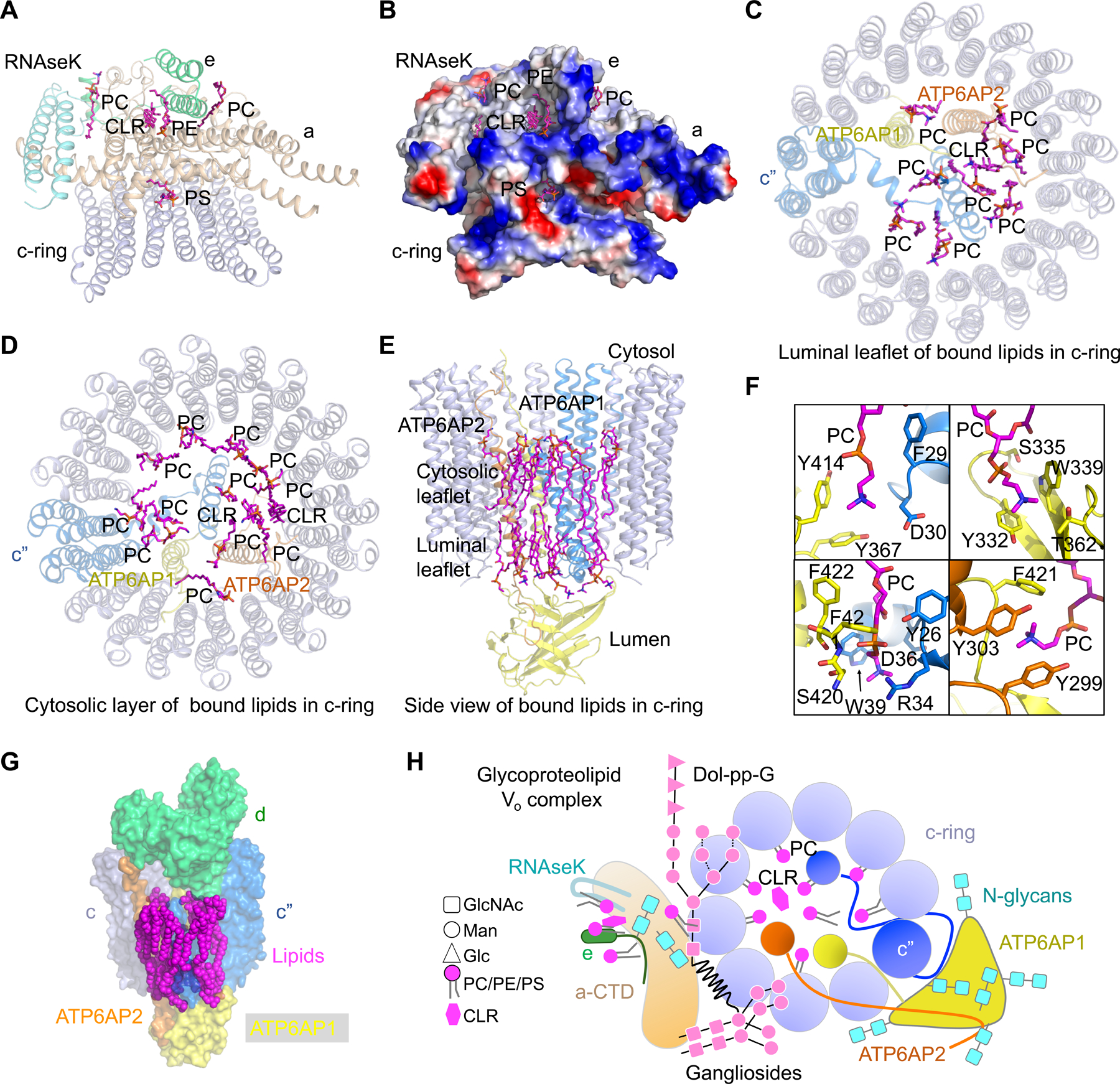
Figure 6. Lipid Molecules in the Vo Complex.

(A) Lipid molecules at the interfaces of subunits a, e, RNAseK, and the c-ring. These lipids establish extensive interactions with the protein subunits above. PC: phosphatidylcholine; PE: phosphatidylethanolamine; PS: phosphatidylserine; CLR: cholesterol.
(B) Electrostatic surface representation of subunits a, e, RNAseK, and c-ring, showing the interactions between the lipids and protein subunits.
(C, D) Lipid molecules inside the c-ring at the luminal leaflet (C) and the cytosolic leaflet (D). These lipids establish extensive interactions with ATP6AP1, ATP6AP2, and the c-ring.
(E) Side view of bound lipids inside the c-ring.
(F) Detailed interactions of lipid molecules with ATP6AP1 (yellow), c” (blue) and ATP6AP2 (orange).
(G) Surface representation of lipid molecules, c-ring, ATP6AP1, ATP6AP2, and subunit d, showing their interactions.
(H) A schematic diagram of the luminal view of the glycoproteolipid Vo complex, showing all protein subunits, N-linked glycans, glycolipids, and ordered PC and cholesterol lipids inside and outside the c-ring.
See also Figure S6.
