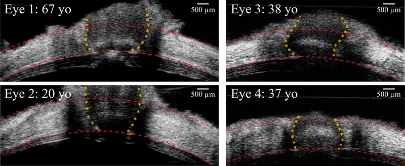
Figure 3:
B-mode images of the posterior eye from four human donors at the initial IOP (5 mm Hg), showing the variations of the ONH morphology. Images on the left column were acquired from Vevo 2100 ultrasound system with an image width of 9.73 mm, and images on the right column were from Vevo 660 with an image width of 8 mm.

