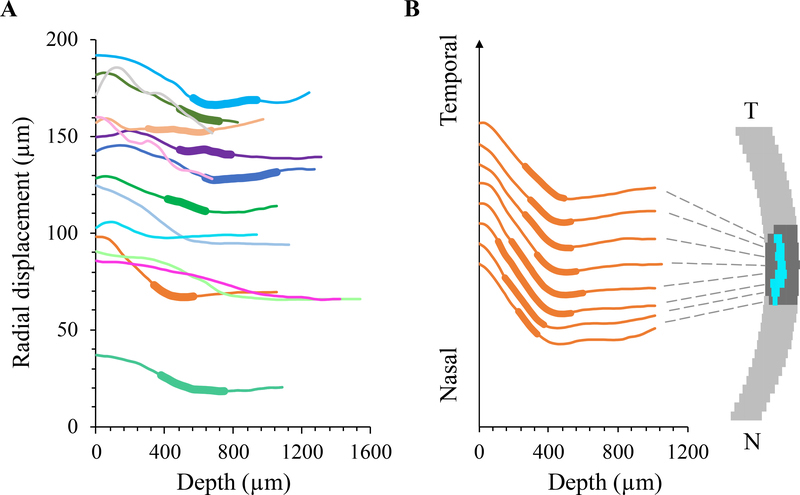
Figure 6:
A. Radial displacements in the center column of the ONH decreased from anterior to posterior ONH with a changing gradient through thickness in each tested eye (n=14, each color represents an individual eye). In eyes whose LC was identified, the LC region was indicated by a thickened segment in the curve. The LC appeared to locate at the transition region where the displacement gradient changed from negative to close the zero. B. Radial displacement curves from several vertical columns of the ONH in a representative eye, showing similar displacement profiles from nasal to temporal ONH. For clearer illustration, the curves were spread vertically by an equal interval at the initial point.

