Figure 7:
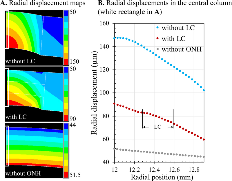
Radial displacements from FE models of human posterior eye simulating a uniform scleral shell (“without ONH”), a scleral shell with an ONH but without LC (“without LC”), and a scleral shell with an ONH and an LC occupying middle third of the thickness of the ONH (“with LC”). A. Radial displacement maps of the posterior eye in all three scenarios. B. Radial displacements in the central column (white rectangle in A) showing that LC significantly reduced the overall displacements of the ONH; however, the simplified models did not reproduce the gradient change in the LC region observed experimentally.

