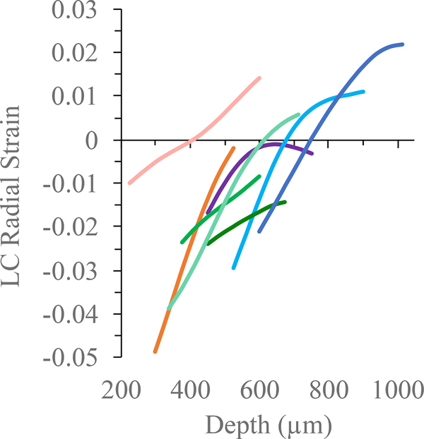
Figure 8:
Radial strain from anterior to posterior LC in the center column of the ONH in eight eyes whose LC was identified (each color represents an individual LC matching the eye’s color in Figure 6). Compressive (negative) strain was highest in the anterior LC and decreased across LC. In four eyes, radial strain became positive in the posterior LC.

