
FIGURE 4. CDM partially support NK cell development.
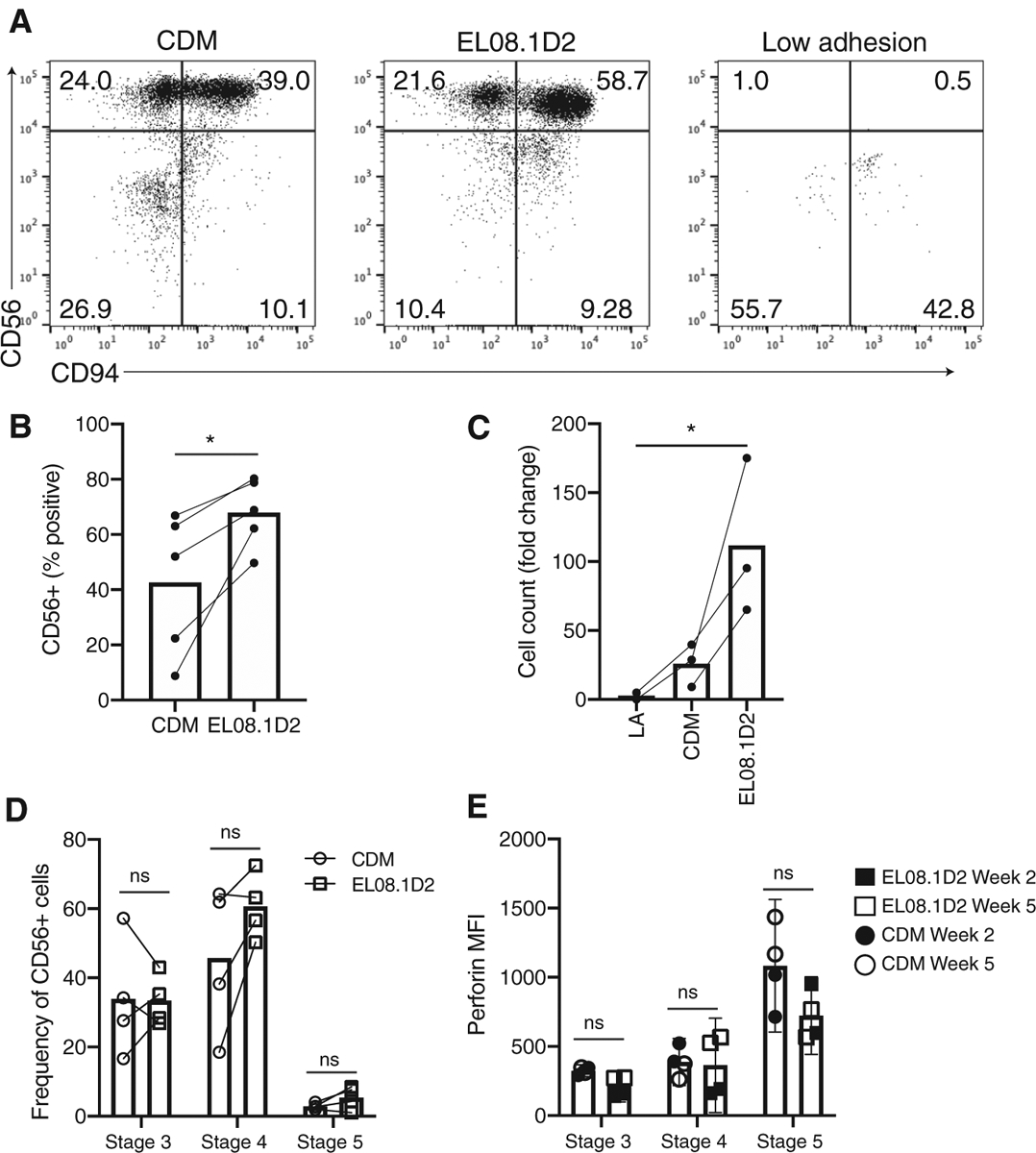
CD34+ HSCs were isolated from peripheral blood and seeded on CDM, EL08.1D2, or low adhesion plates. (A) After 21 days cells were harvested and analyzed by flow cytometry to determine cell maturation. Cells were gated on FSC/SSC and a live-dead marker for live lymphocytes. Representative flow cytometry plots are shown from 2 independent repeats with different donors. (B) Percentage of cells positive for CD94 or CD56 following culture of CD34+ precursors on paired CDM and EL08.1D2 conditions. n = 4 (CD94), 5 (CD56). *P < 0.05 by 2-tailed paired t-test. (C) Cell expansion shown as fold change from number of CD34+ precursors seeded at day 0. n = 2 (low adhesion), 3 (CDM and EL08.1D2) independent replicates from different healthy donors. *P < 0.05 by ordinary 1-way ANOVA with multiple comparisons. (D) Frequency of CD56+ cells in NK cell developmental stage 3 (CD56+CD117+CD94−), stage 4 (CD56+CD94+CD16−), or stage 5 (CD56+CD94+/−CD16+) at day 21 of differentiation. (E) Expression of perforin in mature NK cells differentiated on CDM or EL08.1D2. Cells were gated on stage 3 (CD56+CD117+CD94−) stage 4 (CD56+CD94+CD16−) or stage 5 (CD56+CD94+/−CD16+). n = 4 biological replicates analyzed at day 14 (closed symbols) or day 35 (open symbols) of culture. ns = not significant by two-way ANOVA with Sidak’s multiple comparisons test