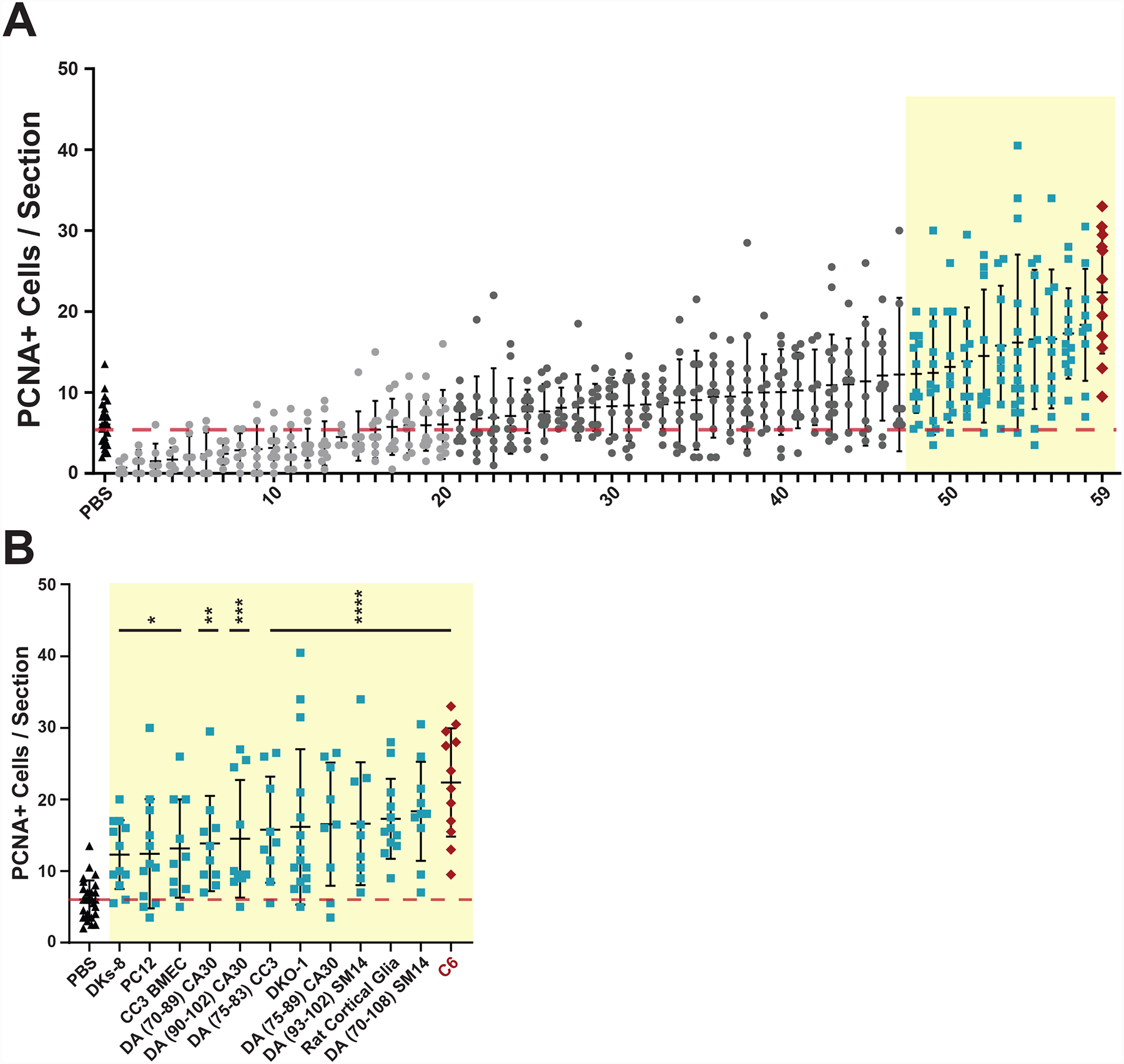
Fig 1. In vivo screen to identify EV sources capable of inducing increased numbers of PCNA+ cells.

EVs were isolated from conditioned media or dissociated tissues and intravitreally injected into undamaged wild type AB zebrafish eyes. After 72 hours, retinas were dissected, sectioned, and immunostained with antibodies against Proliferating Cell Nuclear Antigen (PCNA). A) 59 independent EV preparations were tested and PCNA+ cells were counted across the inner nuclear layer (INL) and outer nuclear layer (ONL) and compared to control PBS injections. Each data point represents PCNA+ cells from a single retina and consists of average counts from 2–4 nonconsecutive sections from the same eye. Light gray EV samples (1–20) led to PCNA counts less than or equal to PBS control background levels (red dotted line). Dark gray EV samples (21–47) induced non-significant PCNA counts slightly greater than background. Light blue EV samples (48–58) induced significant (p-values <0.05) PCNA counts greater than control PBS injections. C6 EVs (red diamonds) induced the most significant PCNA+ counts compared to PBS controls. One-way ANOVA with Dunnett multiple comparison tests (to PBS injection) were used to determine significance. The identify of each EV preparation can be found in Table S1. B Enlargement of EV preparations from (A) that produced significant increases in PCNA counts compared to PBS controls with the indicated source of EVs shown along the X axis. *p-value <0.05, **p-value = 0.0034, ***p-value = 0.0008, ****p-value <0.0001.
