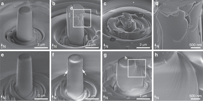
Fig. 4. Deformation of biogenic and geological calcite via uniaxial micro-pillar compression tests.
SEM images of a–d biogenic and e–h geological calcite under sequential deformation steps, including the calcite a, e before compression, b, f immediately after yielding, and c after complete fracturing or g significant plastic deformation. White arrows in f show the initial slip-induced deformation in geological calcite. d, h High-magnification SEM images show d microcracking in the A. rigida biogenic calcite, and h slips in the geological calcite, respectively.

