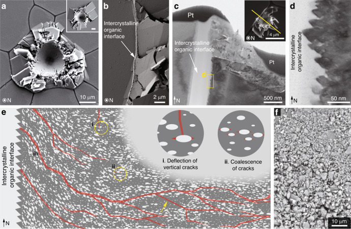
Fig. 7. Synergistic toughening mechanisms across multiple length scales.
a Plan view SEM image of a high-load indentation on A. rigida biogenic calcite (conospherical tip; tip radius = 5 μm; semi-angle = 45°; maximum load = 500 mN). Inset: Plan view SEM image of geological calcite under the same loading condition; scale bar 10 μm. b Damage localization by the intercrystalline organic interfaces as the micro-fracturing in the prism on the right was completely stopped by the interface. c Cross-sectional TEM image showing that the inelastic deformation was effectively localized by the organic interface beneath the sample surface. Inset: Plan view SEM image of the original indentation residue. The yellow solid line indicates the location and orientation of the TEM sample prepared by FIB. Pt, protective platinum layer. d A characteristic saw-tooth-like microstructure observed on the vertical surface of the prisms. e Schematic illustrating the regulation of crack formation and propagation (indicated by red lines) in the A. rigida biogenic calcite due to the intracrystalline defects, including (i) crack deflection; (ii) crack initiation resulting from micro-crack coalescence in regions with high defect density; and (iii) extensive upwardly directed cracks due to the inclined defects near prism boundaries. The yellow arrow shows that regions with a low density of defects fracture similar to pure calcite. f Micro-/nano-scale fractured pieces of the A. rigida biogenic calcite after extensive deformation.

