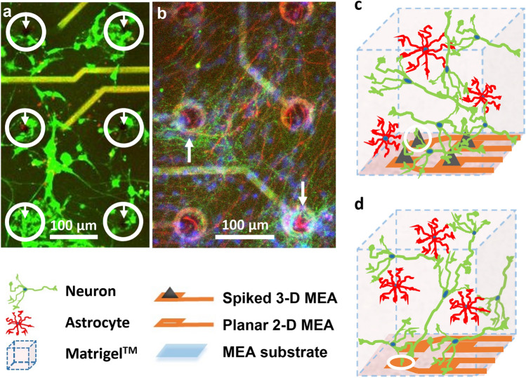
Fig. 2.
Cells and electrodes are closely apposed at the 3-D culture electrode array interface. a Live cells (fluorescing green by AM-cleavage) and dead cells with compromised membranes (nuclei fluorescing red by EthD-1 binding to DNA) are seen in a confocal z-stack image projection of a 105 μm thick region of a 3-D co-culture on a spiked MEA at 24 days in vitro. The co-cultures maintained significant viability for more than 3 weeks in culture, with cells showing in vivo-like somatic shapes and networking via process outgrowth as well as cluster formation. Cells within the white circles are seen apposed around the 3-D electrode spikes demarked by white arrows. b Neurons, astrocytes, and cellular nuclei are seen immuno-stained green (Tau 5), red (GFAP), and blue (Hoechst), respectively, showing random distribution of the two cell types and the apposition of approximately 5–10 cells per electrode in a confocal z-stack image projection of a 80 μm thick culture region on a MEA at 22 days in vitro as indicated by white arrows. This indicates that each electrode likely records activity from multiple cells. c Schematic of the 3-D neuron-astrocyte co-culture interfaced to the spiked (3-D) MEA. d Schematic of the 3-D co-culture interfaced with planar (2-D) MEA. The planar MEAs served as a control for the spiked MEA. In c and d, neurons are depicted in green and astrocytes in red and scale is exploded for illustration. (Color figure online)

