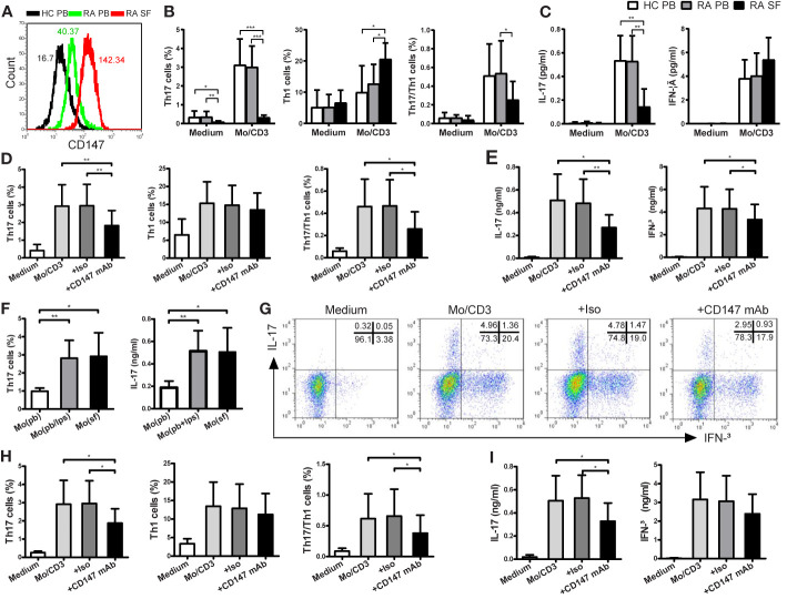
Figure 4.
CD147 suppresses Th17 responses in patients with RA. (A) Representative CD147 staining histogram in CD4+ Tm cells from the peripheral blood of HC (HC PB), PB of RA (RA PB) and synovial fluid of RA (RA SF). (B, C) CD4+ Tm cells from HC PB (n = 13), RA PB (n = 12) or RA SF (n = 6) were isolated and cocultured with medium or autologous PB LPS-activated Mo and anti-CD3 mAb. (D, E) CD4+ Tm cells from RA PB were cocultured with medium or LPS-activated Mo in absence or presence of Iso or anti-CD147 mAb. Percentages of Th17, Th1 and Th17/Th1 cells (B, D) and levels of IL-17 and IFN-γ (C, E) are shown. (F) CD4+ Tm cells from RA PB were cocultured with autologous Mo from PB (Mo (pb)), LPS-activated Mo from PB (Mo (pb/lps)) or Mo from SF (Mo (sf)), then Th17 cell percentage and IL-17 level are shown (n = 6). (G–I) CD4+ Tm cells from RA PB were cocultured with medium or Mo from SF in absence or presence of Iso or anti-CD147 mAb. Representative plots (G) and quantitative results of intracellular expression of IL-17 and/or IFN-γ (H) in T cells, and IL-17 and IFN-γ levels in cell culture supernatants (I) were shown (n = 6). *P < 0.05; **P < 0.01; ***P < 0.001.

