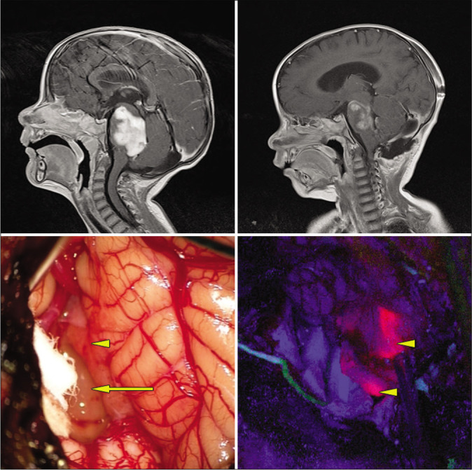
Figure 1:

Exophytic pontine pilocytic astrocytoma. Top left: preoperative, postgadolinium-enhanced sagittal MRI. Top right: postoperative gadolinium-enhanced MRI revealing subtotal resection. Bottom left: intraoperative white light microscopic view. Arrow: obvious tumor under white light microscopy. Arrowhead: normal appearing tissue under white light microscopy. Bottom right: arrow heads: blue light mode reveals solid fluorescence of both obvious tumor and normal appearing area under white light mode.
