Figure 1.
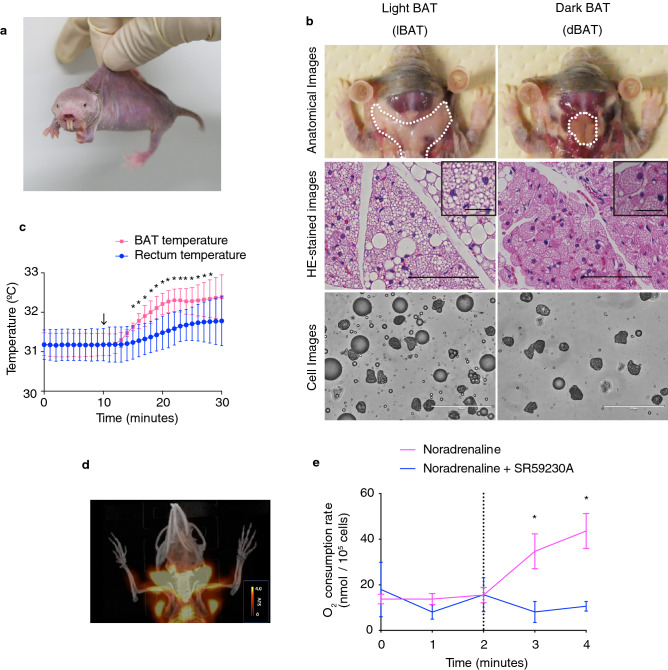
Heterothermic naked mole-rats (NMRs; Heterocephalus glaber) possess thermogenic brown adipose tissue (BAT). (a) Photograph of an adult NMR. (b) Anatomical (top), hematoxylin–eosin (HE)-stained (middle) and cell (bottom) images of light BAT (lBAT) and dark BAT (dBAT). Scale bar = 100 µm for HE-stained images, 25 µm for insets, 200 µm for cell images. (c) BAT and rectum temperatures of anesthetized NMRs before and after the i.p. injection of 1 mg/kg noradrenaline (arrow) at 30 °C (n = 3 animals). *p < 0.05 significantly different from each temperature at 9 min (paired t-test). (d) Positron emission tomography/computed tomography (PET/CT) imaging of NMR-BAT after the injection of 1 mg/kg noradrenaline and 11 MBq 2-deoxy-2-[18F]fluoro-d-glucose ([18F]FDG) at 32 °C. (e) In vitro oxygen consumption rates of isolated adipocytes after the injection of 1 µM noradrenaline (dotted line) with or without pre-incubation with 10 µM SR59230A (n = 3 animals per treatment). *p < 0.05 significantly different from SR59230A treated cells (paired t-test). All data are presented as means ± SEM with the exception of (c), which are means ± SD.

