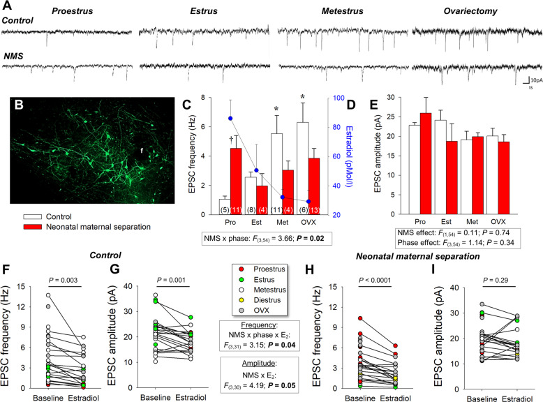
Fig. 5. Neonatal maternal separation (NMS) reduces the excitatory postsynaptic currents (EPSC) recorded in GFP-labeled orexin neurons in response to natural and ovariectomy-induced changes in 17β-estradiol (E2) level.
A Comparison of EPSC recordings from orexin neurons between cells obtained from females during different phase of the estrus cycle and 2 weeks following ovariectomy (OVX); tissue slices originated from females raised under control conditions (top traces) or subjected to neonatal maternal separation (NMS, bottom traces; 3 h/day, postnatal days 3–12). B Photomicrograph illustrating GFP-labeled orexin neurons; the fornix (f) is shown as a landmark. C Population data of EPSC frequencies recorded during three distinct phases of the estrus cycle and following OVX. D Baseline E2 values from Fig. 2A measured in each stage are reported for comparison; values from NMS and controls were pooled since they are not statistically different. E Reports EPSC amplitudes. Note that since recordings in diestrus were performed only in controls (see F, G), data for this stage could not be compared between groups. Effect of E2 application (10 min; 100 nM) on EPSC F frequency and G amplitude in slices from control females. EPSC frequency and amplitude data from NMS females are reported in (H, I), respectively. Within histogram bars, the numbers in brackets indicate the number of replicates in each group. Data are expressed as the mean ± SEM. Post hoc pairwise comparisons were performed only when warranted by ANOVA. †Significantly different from corresponding control value at P ≤ 0.05. *Indicates a value statistically different from corresponding proestrus value at P < 0.05.

