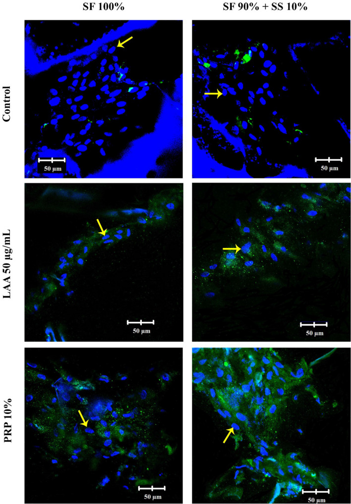
Figure 13.
Immunocytochemistry (ICC) of collagen type II in hWJ-MSCs grown on silk fibroin-spidroin mix scaffold (SF 90% + SS 10%) and silk fibroin scaffold (SF 100%), cultured on differentiation medium with FBS 10% (control), LAA 50 µg/mL, and PRP 10%. The images were observed under confocal microscope after 21 days of culture. Collagen type II appeared green on the images. Yellow arrows indicate the cell nuclei. SF = silk fibroin; SS = silk spidroin.

