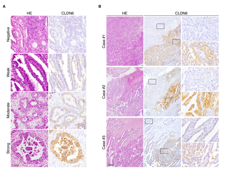
Figure 2.
Immunohistochemical staining of CLDN6 in endometrial cancer tissues. (A) Representative immunohistological images showing negative/weak/moderate/strong SI for CLDN6 expression in endometrial cancer tissues. HE, hematoxylin–eosin. Scale bar, 50 mm. (B) Intratumoral heterogeneity of CLDN6 protein in the subjects with endometrial cancer with high CLDN6 expression. The rectangles indicate CLDN6-positive and -negative subpopulations. Scale bar, 200 µm.

