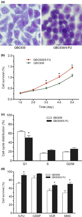
Figure 1.

Biological characteristics of QBC939/5‐FU cells. (a) Cellular morphology dyed with 0.005% crystal violet for 30 min. Zoom: 200×. (b) Growth curve assayed by MTT. (c) Cell cycle distribution determined by flow cytometry. (d) Effects of four common chemotherapeutics (60 μM, 24 h) on the proliferation of QBC939 and QBC939/5‐FU cells assayed by MTT. *P < 0.05, **P < 0.01. 5‐FU, 5‐fluorouracil; CDDP, cis‐diammineplatinum(II) dichloride; VCR, vincristine sulfate; MMC, mitogen enzyme C.
