Figure 4.

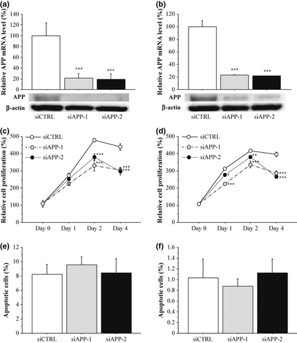
Effects of amyloid precursor protein (APP) on cell proliferation and apoptosis in breast carcinoma cells. (a, b) Expression of APP in MCF‐7 (a) and MDA‐MB‐231 (b) cells transfected with APP‐specific siRNA (siAPP‐1 and siAPP‐2) or negative control siRNA (siCTRL) at mRNA level evaluated by real‐time PCR (upper panels) and protein level by immunoblotting (lower panels). For the immunoblotting, 10 μg of protein was loaded in each lane, and β‐actin immunoreactivity is shown as the internal control. (c, d) Proliferation activity of MCF‐7 (c) and MDA‐MB‐231 (d) cells transfected with siRNA summarized as a ratio (%) compared to that 0 days after treatment. (e, f) Percentage of apoptotic cells in the MCF‐7 (e) and MDA‐MB‐231 (f). Open bar/circle: siCTRL, gray bar/circle: siAPP‐1; and closed bar/circle: siAPP‐2. Data were presented as the mean ± SD (n = 3), respectively. *P < 0.05, **P < 0.01 and ***P < 0.001 compared with siCTRL groups.
