Figure 3.
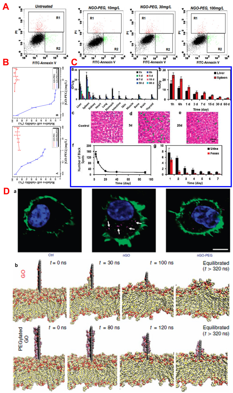
(A) Apoptosis assay was done by FACS. MCF-7 cells treated with different concentrations of NGO-PEG. R1 (red) represent region of dead cells and R2 (green) represent the regions of apoptotic cells. Reprinted with permission from Liu Z, Robinson JT, Sun X, Dai H. PEGylated nanographene oxide for delivery of water-insoluble cancer drugs. J Am Chem Soc. 2008;130(33):10876-10877.Copyright 2008 American Chemical Society.11 (B) Cell viability of GO-PEG (red line) and GO-PEG/PTX against A549 (A) and MCF-7 (B) after 72 h of incubation. Reprinted with permission from Xu Z, Wang S, Li Y, Wang M, Shi P, Huang X. Covalent functionalization of grapheneoxide with biocompatible poly (ethylene glycol) for delivery of paclitaxel. ACS Appl MaterInterfaces. 2014;6(19):17268-76. Copyright 2014 American Chemical Society.10 (C) In vivo biodistribution and clearance data of NGS-PEG in female Balb/cmice. (a) Time-dependent biodistributionin major organs of 125I-NGS-PEG. (b) presence of 125I-NGS-PEG in the liver and spleen over different time points. (c-e) Haemotoxylin and Eosin (H&E) stained liver sections from the untreated control mice(c) and NGS-PEG treated mice at day 3 (d) and day 20 (e) of post injection. (f) Statistic of the numbers of black spot in liver sections at various time point after NGS-PEG administration. Each data point was the average of 5 images under a 20X objective. (g) the levels of 125I-NGS-PEG in urine and faeces in the first week of post injection. Excretions of mice were collected by metabolism cages. Standard deviations of 4-5 mice per group were taken for the error bar. Reprinted with permission from Yang K, Wan J, Zhang S, Zhang Y, Lee ST, Liu Z. In vivo pharmacokinetics, long-term biodistribution, and toxicology of PEGylated graphene in mice. ACS Nano. 2011;5(1):516-22. Copyright 2011 American Chemical Society13 and (D) Internalization of nGO, nGO-PEG by peritoneal macrophages (white arrow indicated the nGO as purple dots) (a), Snapshots of membrane insertion processes of GO and PEGylated GO during stimulation, (carbons of GO represented in grey color and covalently linked PEG chains are in purple color) (b). Reproduced from Luo N, Weber JK, Wang S, et al. PEGylated graphene oxide elicits strong immunological responses despite surfacepassivation. Nat Commun. 2017;8(1):1. Creative Commons license and disclaimer available from: http://creativecommons.org/licenses/by/4.0/legalcode.47

