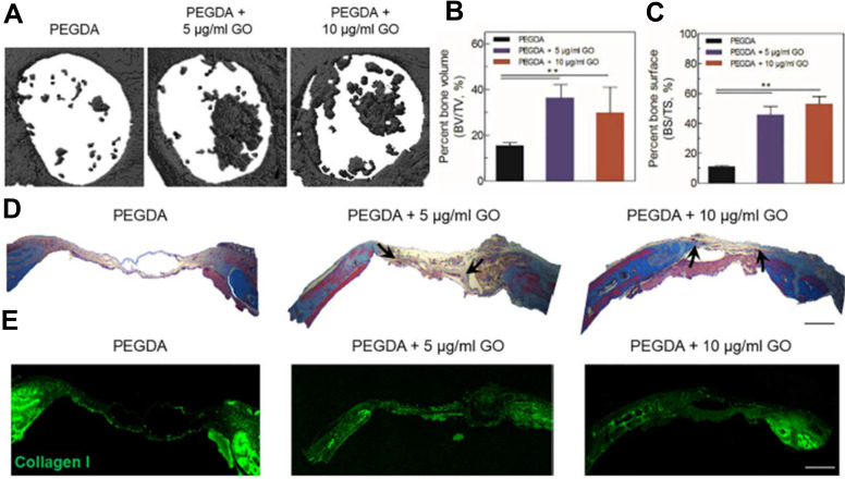
Figure 6.
(A) Micro-CT image of new bone formation in mouse calvaria defect model (defect dia. = 4 mm). (B) Percent bone volume and (C) percent bone surface of PEGDA + 5 μg/mL GO and PEGDA + 10 μg/mL GO group was significantly higher than hTMSCs on PEGDA group (n = 3, p < 0.01). (D) Masson’s trichrome staining for histological analysis of regenerated bone tissue (scale bar = 500 µm). (Black arrow indicated the newly deposited collagen) (E) Immunostaining was done for collagen type I in regenerated bone for each groups (scale bar = 500 µm). Reprinted with permission from Kim HD, Kim J, Koh RH, et al. Enhanced Osteogenic commitment of human Mesenchymal stem cells on polyethylene glycol based Cryogel with Graphene oxide substrate. ACS Biomater Sci Eng. 2017;3(10):2470-2479. Copyright 2017 American Chemical Society.64

