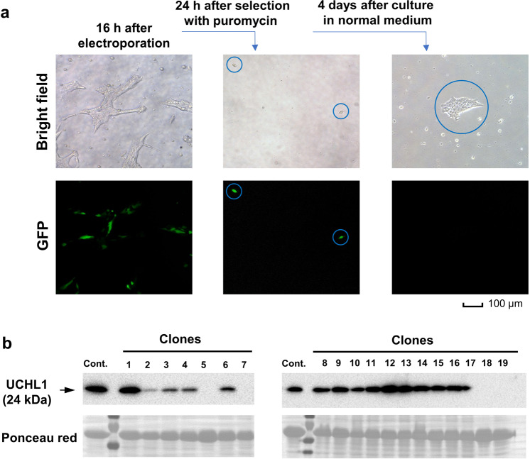
Fig. 3. Early procedures for electroporation, puromycin selection, and analysis of UCHL-1 expression by immunoblotting.
a Representative bright-field (upper panels) and GFP (lower panels) microscope images at the indicated time points after electroporation. b Single colonies of edited cells were analyzed by immunoblotting for the expression of UCHL-1. Ponceau red staining was used to indicate equal loading.

