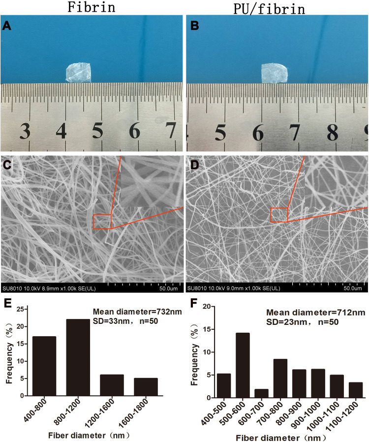
Figure 1.
Morphological observation and fiber diameter analysis of electrospun vascular scaffolds. The macroscopic morphology of fibrin (A) and PU/fibrin (15:85) (B) scaffolds was collected with the camera. Microstructures of fibrin (C) and PU/fibrin (15:85) (D) vascular scaffolds were observed by SEM. The fiber diameter distribution of fibrin (E) and PU/fibrin (15:85) (F) were measured by ImageJ software (n=3).

