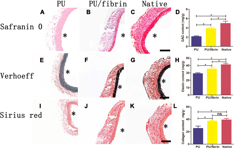
Figure 8.
Histological characteristics of ECM changes in PU/fibrin (15:85) vascular graft at 3 months. The samples were stained with Safranin O staining reagent for glycosaminoglycans (A–C), Verhoeff staining reagent for elastin (E–G), and Sirius red staining reagent for collagen (I–K). Scale bar: 100μm. Quantitative analysis of GAG (D), elastin (H) and collagen (L) content in vascular grafts (n=3). * indicated the graft lumen. ★:P < 0.05, ns = not significant.

