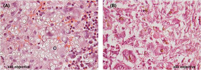
Figure 5.
Histology images for fungal infections: (A) African histoplasmosis; Hematoxylin and eosin (H&E) stained sections showing multinucleate giant cells containing numerous large yeast cells in their cytoplasm. The yeast cells are approximate 10–15um in diameter and are thick walled. (B) Chromoblastomycosis; H&E stained sections showing pigmented, brown, round organisms with thick walls ‘Copper bodies’. This Figure is reproduced in color in the online version of Medical Mycology.

