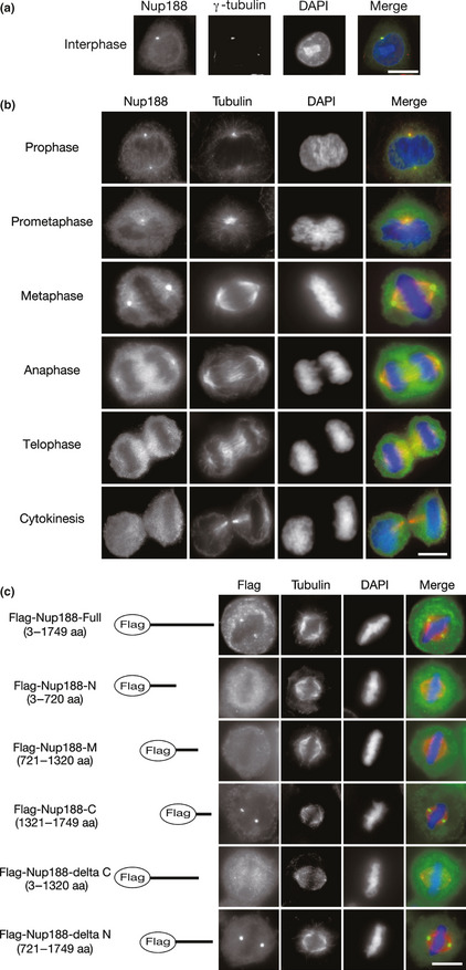
Figure 1.

Nup188 localizes to centrosomes in mitosis. (a) Subcellular localization of Nup188 in interphase. HeLa cells were stained with anti‐Nup188 (green) and anti‐γ‐tubulin (red) antibodies. DNA was stained with DAPI (blue). Bar, 10 μm. (b) Subcellular localization of Nup188 during mitosis. HeLa cells were stained with anti‐Nup188 (green) and anti‐α‐tubulin (red) antibodies. DNA was stained with DAPI (blue). Bar, 10 μm. (c) Subcellular localization of Nup188 deletion mutants in metaphase. Localization of Flag‐tagged full‐length Nup188 or deletion mutants expressed in HeLa cells is shown (green). Microtubules were stained with anti‐α‐tubulin antibody (red) and DNA was stained with DAPI (blue). Bar, 10 μm.
