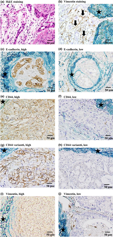
Figure 3.

Immunohistochemical staining of tumor tissue in patients with lung adenocarcinoma with intratumoral vascular invasion. Stars indicate elastic fibers with positive Victoria blue van Gieson staining. (a,b) Intravascular stromal cells stained with (a) H&E and (b) immunohistochemical staining for vimentin. The arrows point to round or spindle‐shaped, brown‐stained, non‐cancerous cells. The staining results were negative for cancer cells. (c,d) E‐cadherin expression in cancer cells. (e,f) CD44 expression in cancer cells. (g,h) CD44 variant 6 expression in cancer cells. (i,j) Vimentin expression in cancer cells.
