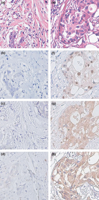
Figure 1.

Representative photographs of androgen receptor (AR), 5α‐reductase type 1 (5αR1), and 17β‐hydroxysteroid dehydrogenase type 5 (17βHSD5) immunohistochemistry in AR−5αR1−17βHSD5− (a–d) and AR+5αR1+17βHSD5+ (e–h) triple negative breast carcinomas. Hematoxylin–eosin staining (a,e). Androgen receptor was immunolocalized in the nuclei of carcinoma cells at variable immunoreactivity (b,f). Both 5αR1 (c,g) and 17βHSD5 (d,h) were immunolocalized in the cytoplasm of carcinoma cells. Stromal cells were negative in areas either adjacent or distal to carcinoma. Original magnification, x200.
