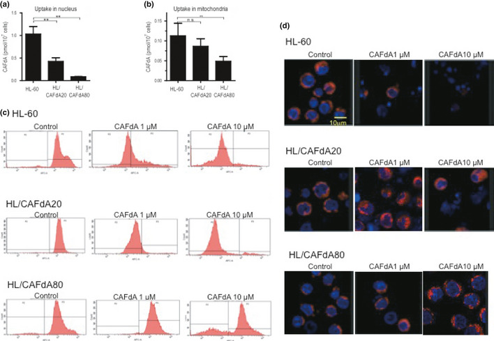
Figure 3.

(a,b) The cells (HL‐60, HL/CAFdA20 and HL/CAFdA80) were incubated with tritiated CAFdA at 0.2 μM for 24 h, and fractionated into cytoplasm, mitochondria and nuclei, followed by scintillation counting of the radioactivity of each fraction. Data shown are the means and SD of three independent experiments. (c) CAFdA‐mediated mitochondrial damage. Mitochondrial membrane potentials were determined using near infrared (NIR) stain treated with 1 or 10 μM CAFdA for 24 h. Representative flow cytometry is shown. (d) Double stains were also performed in the cells at 24 h after administration of 1 or 10 μM CAFdA. Nuclei were dyed by Hoechst 33342, while mitochondria were dyed by NIR.
