Figure 4.

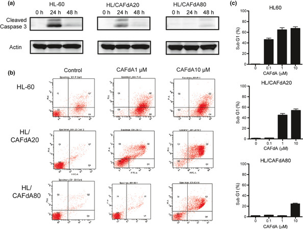
Induction of apoptosis. (a) Time course of cleaved caspase 3. Cleaved caspase 3 was detected in the cells at 24 and 48 h after 1 μM CAFdA treatment. Data are representative of at least three biological replicates. (b) The induction of apoptosis detected after 24‐h incubation with CAFdA (1 and 10 μM), using annexin V and propidium iodide stain. Annexin V‐positive cells are considered apoptotic. Data are representative of at least three replicates. (c) The ratio of sub‐G1 was determined by flowcytometric analysis in the cells after 24‐h incubation with CAFdA (0.1, 1 and 10 μM).
