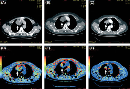
Figure 1.

Imaging of the contrast computed tomography (CT) scan and perfusion of a case treated with combined Navelbine and platinum (NP) and Re‐endostatin. (A) Contrast CT image before treatment. (B) Contrast CT image after two cycles of treatment without tumor shrinkage (arrow). (C) Contrast CT image after four cycles of treatment with tumor shrinkage (arrow). (D) Blood volume inside the tumor before treatment (arrow). (E) Decreased blood volume inside the tumor (arrow) after two cycles of treatment. (F) Continuously decreased blood volume inside the tumor after four cycles of treatment (arrow).
