Figure 2.

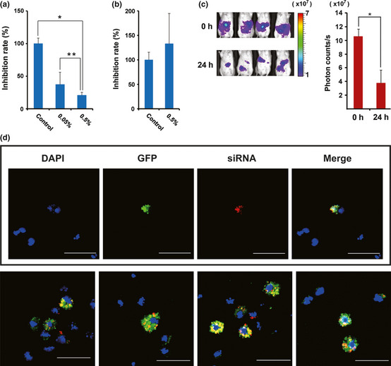
Evaluation of an atelocollagen‐mediated siRNA delivery system by measuring the luciferase activity after i.p injection of luciferase siRNA. (a) Inhibition rates of photon counts 48 h after injection are compared between 0.05% and 0.5% atelocollagen/luciferase siRNA/DharmaFECT1. *P = 0.006; **P = 0.012. (b) Inhibition rates of photon counts 48 h after injection are compared between 0.5% atelocollagen/control siRNA and 0.5% atelocollagen/luciferase siRNA. No significant inhibition is observed in a luciferase siRNA complex containing no DharmaFECT1. (c) Reduction of luciferase activity 24 h after injection with 0.5% atelocollagen/luciferase siRNA/DharmaFECT1 is visualized in four representative mice (left). Significance of the reduction is also shown (right). *P < 0.0001. Color bar indicates × 107 photon/s. (d) Delivery evidence of siRNA to cancer cells in the peritoneal cavity. Most green fluorescent protein (GFP)‐expressing 60As6 cells (green) incorporate fluorescence‐labeled siRNA (red). Bar, 50 μm.
