
Figure 3.
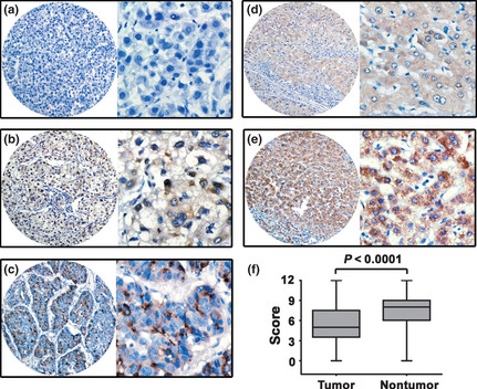
Expression of cyclin F was decreased in hepatocellular carcinoma (HCC) tissues by immunohistochemistry. Cyclin F was presented predominantly in cytoplasm within tumor and normal liver cells. The micrographs showed negative (a), weak (b) and strong (c) staining of cyclin F in HCC, as well as weak (d) and strong (e) staining of cyclin F in normal liver tissues. (Left panel: magnification ×100; right panel: magnification ×400.) (f) Reproducibility of the measurement in all 245 patients was calculated using the Wilcoxon matched paired test.