
Figure 3.
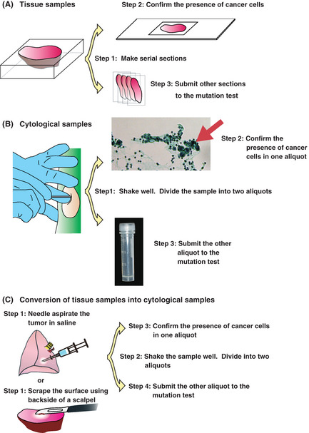
Sample preparation procedures. (A) Tissue samples. Step 1: Serial sectioning. Step 2: The presence of cancer cells is confirmed in 1 section. Step 3: The EGFR mutation is investigated using other sections. Macro‐dissection may be required to remove normal tissue before step 1. (B) Cytological samples. Step 1: Suspend the cells in saline. Divide the samples into two aliquots. Step 2: Confirm the presence of cancer cells in one aliquot. Step 3: Investigate the EGFR mutation using the other aliquot. (C) Preparation of cytological samples from tissue. Step 1: Scrape the surface of the tissue. Suspend the cells in saline. Step 2: Divide the samples into two aliquots. Step 3: Confirm the presence of cancer cells in one aliquot. Step 4: Investigate the EGFR mutation using the other aliquot.