Figure 9.

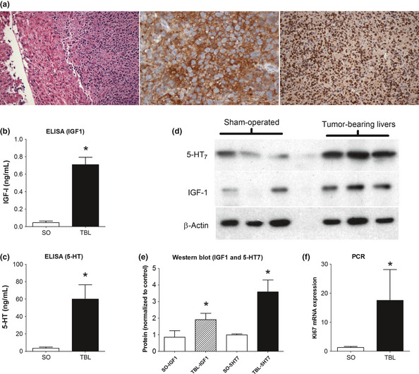
IGF‐1 and 5‐HT7 in a murine model of hepatic SI NEN metastasis. Nude mice injected splenically with H‐STS cells develop liver metastases within 4–6 weeks. Tumors demonstrate typical NEN morphology (H&E, left), are CgA positive (DAB‐brown stained cells, center) and are rapidly proliferating (Ki67: 90%, right) (a). IGF‐1 (b) and 5‐HT (c) were significantly increased in tumor‐bearing livers compared to sham‐operated animals. Western blot confirmed elevated 5‐HT7 and IGF‐1 in TBL animals (d); optical density (ImageJ software) confirmed over‐expression compared to SO mice (e). Ki67 mRNA expression was increased approximately 15‐fold compared to SO mice. SO, sham‐operated; TBL, tumor‐bearing livers; mean ± SEM, n = 6–9.
