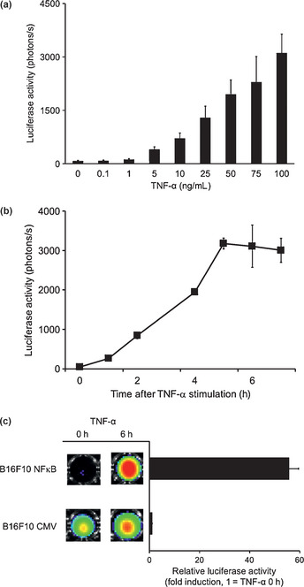
Figure 1.

Nuclear factor‐κB (NFκB) activation after tumor necrosis factor‐α (TNF‐α) treatment in B16F10 mouse melanoma cells. (a) B16F10 NFκB cells were incubated with indicated concentrations of TNF‐α for 6 h and the luminescence was measured. (b) B16F10 NFκB cells were treated with TNF‐α (100 ng/mL) and the luminescence was measured at the indicated time after the TNF‐α stimulation. (c) B16F10 NFκB and B16F10 CMV cells were stimulated TNF‐α (100 ng/mL) for 6 h. The luminescence was measured at 0 h and 6 h after TNF‐α stimulation. Error bars represent SEM.
