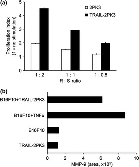
Figure 3.

Functional roles of the tumor necrosis factor‐related apoptosis‐inducing ligand (TRAIL)–DR5 pathway in B16F10 mouse melanoma cells. (a) B16F10 CMV cells were cocultured with TRAIL‐2PK3 or control 2PK3 cells at responder (R):stimulator (S) ratio of 1:1 for 48 h and luminescence was measured. Error bars represent SEM. (b) B16F10 cells were stimulated with tumor necrosis factor‐α (TNF‐α; 50 ng/mL) or cocultured with TRAIL‐2PK3 (at R:S 1:1) for 48 h, and the cell‐free supernatant was collected. Gelatin zymography was used to determine MMP‐9 production and the band intensity was measured.
