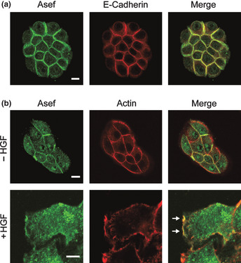
Figure 1.

Subcellular localization of Asef/Asef2 in MDCK II cells. (a) Subcellular localization of Asef/Asef2 and E‐cadherin in small colonies of MDCK II cells. (b) Subcellular localization of Asef/Asef2 in MDCK II cells treated with HGF. After stimulation with HGF (20 ng/mL for 2 h), cells were stained for Asef/Asef2 and actin (trimetylrhodamine isothiocyanate [TRITC]‐phalloidin). Arrows indicate the regions of Asef/Asef2 accumulation in membrane ruffles. Bar, 10 μm.
