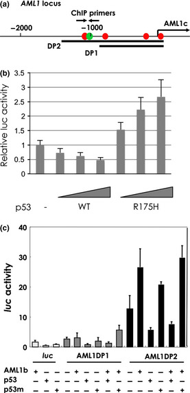
Figure 4.

p53 inactivates AML1 promoter. (a) The indicated promoter regions of 900 bp (DP1) and 1400 bp (DP2) were cloned into luc reporter plasmids. The arrow labeled AML1c indicates the transcription start site. The opposing arrows indicate the positions of the primers used for ChIP analysis. (b, c) AML1 promoter is repressed by p53. SaOs2 cells were transfected with expression vector for wild‐type p53, p53R175H and/or AML1b together with a human AML1 distal promoter–luciferase (luc) reporter containing DP1 or DP2. Data shown are relative values of firefly luciferase activity normalized to Renilla luciferase activity. The data shown are the means ± standard error of the mean (SEM).
